

Main Main principles Neck examination with CT scan: Body Balance Clinic method
BODY BALANCE CLINIC
”BODY BALANCE"
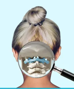
Restoration of the joints of the head
Restoring body biomechanics
Endomassage

FOR PATIENTS
Self-diagnosis
Surveys
Recommendations
Make an appointment
FOR MEDIA

Cooperation