

Main Main principles Impact on the nervous system
BODY BALANCE CLINIC
”BODY BALANCE"
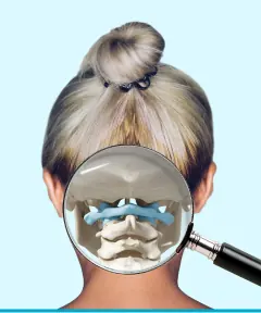
Restoration of the joints of the head
Restoring body biomechanics
Endomassage
FOR PATIENTS
Self-diagnosis
Surveys
Recommendations
Make an appointment
FOR MEDIA
Cooperation