MENU
  • search
  • en

Home Main principles Biomechanical features

Biomechanical features of the craniovertebral region

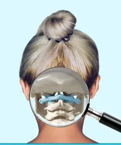
The first two cervical vertebrae (Atlas and Axis), together with the occipital bone of the skull, form 3 head joints: the paired atlanto-occipital joint, the paired lateral atlanto-axial joint and the median atlanto-axial joint of Cruveilhier. In medicine, this region is called the “craniovertebral junction” (from Latin Cranium – skull, from Latin Vertebrae – vertebrae).
These joints balance the center of gravity of the head and equilibrate it on the four sides of the skeleton along the axis of the spinal column, thus influencing the center of gravity of the whole body. The joints provide mobility of the head, protect the spinal cord and are a powerful reflexogenic zone.
In addition to the occipital bone of the skull and the first two cervical vertebrae, the craniovertebral region also includes the medulla oblongata and the cerebellum.

Atlanto-occipital joint

Biomechanical features of the craniovertebral region
The atlanto-occipital joint is a paired articulation located between the occipital bone of the skull and the first cervical vertebra, the Atlas. The joint is formed by the two condyles of the occipital bone of the skull and the two superior articular surfaces of the atlas. Both pairs of articulating surfaces are enclosed in joint capsules.
The following movements are possible in the atlanto-occipital joint:
  • ✅ flexion / extension;
  • ✅ lateral flexion;
  • ✅ combined rotation.

Biomechanics of the atlanto-occipital joint in figures, in normal and pathological conditions

Biomechanical features of the craniovertebral region
1. Flexion / extension – this is bending and straightening the head forward and backward.

Total range of motion in normal conditions: up to 30° (10° flexion, 20° extension). Movement in the joint occurs as a rocking of the skull’s condyles forward and backward on the articular surfaces of the Atlas.
Total range of motion in pathology: down to 0° (0-7.5° flexion, 0-15° extension).
Total range of motion in normal conditions: up to 30° (10° flexion, 20° extension). Movement in the joint occurs as a rocking of the skull’s condyles forward and backward on the articular surfaces of the Atlas.
In pathology, there is a moderate or complete restriction of joint mobility. The patient may have limitation when trying to bend / straighten the head; cannot touch the collarbone with the chin; feels a blocking of the backward extension movement of the head and tension / pain in the thoracic region. When pathology is present, there is often crunching of the articulating surfaces of the joints, shooting pains and an aching pain in the neck that turns sharp at maximum flexion / extension of the head forward / backward.
Biomechanical features of the craniovertebral region
2. Tilting the head left and right / lateral bending (lateral flexion).
Total range of motion in normal conditions: up to 20° (5-10° in each direction).
Sideways tilting in the atlanto-occipital joint occurs through opposite movements of the skull and the vertebra. As the head tilts, a sharp rotation of the vertebra occurs at the same time. The patient’s head tilts to one side, while the vertebra rotates to the other. For example, tilting the head to the right is accompanied by rotation of the atlas to the left.
Total range of motion in pathology: down to 0° (0-7.5° in each direction).
In pathology, there is unilateral partial restriction or complete blocking of joint mobility. The patient may feel that the head tilts worse to one side than to the other. The same movements in different directions may be performed with varying degrees of difficulty, accompanied by painful sensations and shooting pains. With blockage, the articulating surfaces of the joints are subjected to excessive friction, as a result of which crunching and clicking may be heard.
Biomechanical features of the craniovertebral region
3. Combined movement that results in rotation of the head.
Total range of motion in normal conditions: 5-7° in each direction. 
Rotation of the head left and right in the atlanto-occipital joint occurs through opposite movements of the skull and the vertebra. Turning of the head occurs during lateral flexion due to the gliding of the skull along the inclined articular surfaces of the Atlas (their anteromedial orientation) and the alar ligaments. During the tilting and gliding of the skull’s condyles along the articular surfaces of the Atlas, a sharp rotation of the vertebra occurs. 
Total range of motion in pathology: down to 0° (0-5° in each direction).
In pathology, there is unilateral partial restriction or complete blocking of joint mobility. During combined movements, the crunching of the articulating surfaces of the joints, which occurs as a result of the intense compressive force of the muscles, may be especially pronounced or not audible at all (complete block). The patient may feel that the head turns worse to one side than to the other. The same movements in different directions may be performed with varying degrees of difficulty, accompanied by painful sensations and shooting pains.

Lateral atlanto-axial joint

Biomechanical features of the craniovertebral region
The second cervical vertebra, the Axis, differs from all other vertebrae in its unusual shape, namely the presence of the odontoid process, around which the first cervical vertebra, the Atlas, rotates, and the skull along with it.
The lateral atlanto-axial joint is formed by the paired articulation of the C1-C2 vertebrae. It is located between the articular surfaces and processes of the first and second cervical vertebrae, the Atlas and the Axis. In this complex of joints, one single movement takes place — rotation of the head to the right and left.
The lateral atlanto-axial joint provides significant rotation of the head left and right. The normal range of rotational movement is up to 33° to each side out of the 90° of total rotation of the entire cervical spine. In case of joint hypermobility, rotation can reach 40°.
In patients with C1-C2 segment dysfunction who suffer from headaches, rotation may be significantly reduced due to inferior oblique muscle syndrome and, in rare cases, completely blocked. When there are displacements in the vertebrae, a peculiar prolonged rustling of the articular surfaces rubbing against each other may be heard, which the patient usually picks up in silence.

Median atlanto-axial joint of Cruveilhier

Biomechanical features of the craniovertebral region
The median atlanto-axial joint of Cruveilhier is formed by the posterior surface of the anterior arch of the first vertebra and the dens of the second cervical vertebra. 
This is a rather complex joint which, like the lateral atlanto-axial joint, takes part in rotating the head right and left, but simultaneously with this movement, it accounts for flexion-extension of the head of up to 7° in each direction.
Dysfunction and restricted mobility in the Cruveilhier joint can lead to numerous neurological manifestations, especially headaches and migraines.

Testing the mobility of the Cr-C1-C2 segments with the Body Balance Clinic method

Biomechanical features of the craniovertebral region
The cervical spine is divided into 3 parts: upper, middle and lower. 
The head joints belong to the upper cervical segment. They are formed by the Atlas C1 and Axis C2 vertebrae together with the occipital bone of the skull, Cr. When determining dysfunction in these joints, it is not enough to check the patient’s overall head rotation, which is performed with the participation of all three cervical segments.
It is necessary to check, in isolation, the mobility of the upper part of the neck only for the ability to perform various head movements: turns and tilts in different directions.
For this purpose, Body Balance Clinic has developed an individual method for testing the biomechanics of the head joints, which in terms of its accuracy is much more precise than the flexion-rotation test used in kinesiology.
When performing the test, the patient sometimes cannot turn the head at all because of blockage of the head joints.
As you already know, the joints should provide head rotation totalling up to 35-42° to each side. With blockage of these joints, the body compensates for the imbalance by distributing the muscle load onto the trapezius muscles. As a result, a patient with blocked head joints may experience sharp pain even with moderate squeezing of the trapezius muscles, which is not observed if the head joints work correctly.

2 types of head joints dysfunction and their key features

Body Balance Clinic distinguishes 2 types of head joints dysfunction:
  • Type 1 – blockage of the mobility of the head joints by the spasmodic compressive force of the muscles without displacement of the cervical vertebrae;
  • Type 2 – blockage of the movements of the head joints by the spasmodic compressive force of the muscles with displacement of the cervical vertebrae.
Body Balance Clinic’s internal statistics, compiled by analyzing a large number of patients’ CT scans, show that ~ 30% of patients belong to type 1 and ~ 70% of patients to the second type. The second type includes patients who have a rotational or lateral displacement of more than 3° of at least one of the vertebrae and in whom biomechanical tests also reveal blockage of the mobility of the head joints.
Images of serious displacements: 

The condition of the muscles in head joints dysfunction

A muscle can be in 4 different states: normal, contracture, flaccidity and local myofascial hypertonus (a fibrous and painful muscle induration in which the muscle shortens somewhat, which is an indicator of muscle tone).
In head joints dysfunction, the patient’s occipital muscles are almost always in a state of contracture or local myofascial hypertonus (rarely). Body Balance Clinic distinguishes 4 degrees of muscle contracture:
  • 1 first — the muscle is soft or denser than normal, the fingers easily penetrate into its thickness, causing minimal tenderness;
  • 2 second — the muscle is dense, the fingers only partially sink into its thickness, causing moderate or intense tenderness;
  • 3 third — the muscle is of “stony” density and does not let the fingers into its thickness, palpation causes a sensation of severe tenderness;
  • 4 fourth – an inflamed muscle, significantly enlarged compared to its normal size, moderately hard, causes excessive tenderness on palpation.
The long-term persistence of muscle spasms leads to the development of local painful indurations in the muscles (areas of fibrosis), which over time increases their stiffness and the stage of their condition.

Professors of medicine on the importance of the correct function of the head joints

The craniovertebral junction is an important strategic zone that largely determines a person’s health. That is why for many years it has been in the focus of attention of neurologists, vertebrologists, specialists in manual therapy and osteopathy, including cranial osteopathy. Body Balance Clinic strives to structure the available knowledge and publishes some scientific discoveries made by professors of medicine so that the reader can once again be convinced of the importance of the problem and the need for a professional approach to it:
? The craniovertebral junction plays a key role in the redistribution of body weight and in the formation of the supporting function of the lower limbs (Ivanichev G.A., 2008).
?Dysfunction of the craniovertebral junction and the functional block Cr-CI contribute to impaired mobility of the sacrum between the iliac bones, decreased tone of the iliopsoas muscles and the development of twisted pelvis syndrome (Zozulya I.S., Bredikhin A.V., 2011; Chikurov Yu.V., 2004).
?According to G. Goodheart (1968), decreased tone of the iliopsoas muscles is associated with the presence of a Cr-C1 block. Functional blockade of the craniovertebral region is, as a rule, accompanied by secondary blockades of the sacroiliac joint (Ivanichev G.A., 2008).
?The dura mater of the spinal cord is firmly attached to the foramen magnum, the atlas and the axis, so any impairment of mobility in this zone affects the craniosacral mechanism (Orel A.M., 2009).
? Kinetic dysfunctions at this level change the patterns of the sphenobasilar synchondrosis (SBS), which leads to a change in the primary respiratory mechanism (Novoseltsev S.V., 2009).
? In craniovertebral dysfunction, dizziness, more often non-systemic, decreased hearing and hearing loss may be observed. One of the main syndromes is loss of balance with a sudden change in head position, especially when moving from a lying to a standing position (resembles vestibular ataxia) (Ivanichev G.A., 2008).
? Craniovertebral dysfunction may be accompanied by cervicogenic headache, which occurs against the background of impaired spinal biomechanics and myofascial dysfunction with the formation of local muscle spasm and trigger points. Cervicogenic headache can develop through three mechanisms: vascular (vasomotor, ischemic and venous), neuralgic and muscle tension (Morozova O.P., Yaroshevsky A.A., Zdybsky V.I., 2009).
? Reflex spasm when the inferior oblique muscle of the head is stretched, combined with hyperflexion, provokes subluxation of the atlanto-axial joint. In this case, the greater occipital nerve and the occipital artery, which approaches the CI-CII intervertebral joint on the side opposite to the head rotation, may be compressed between the layers of the muscle’s fascia (Bogorodinsky D.K., Skoromets A.A., 2008).
? Impaired craniovertebral biomechanics leads to spasm of the muscles of the head and cervical spine and of the extravertebral muscles, contractures of these muscles, the appearance of latent trigger points in them (muscular, fascial, musculoligamentous, ligamentous-osseous) and regional muscle imbalance. Functional muscular, fascial, musculoligamentous and dural craniovertebral blocks C0-CI, CI-CII develop (Ivanichev G.A., 2008), and irritative-reflex syndromes arise: As a result, the following clinical syndromes form: cervicocranialgia, cervicalgia, cervicobrachialgia, cervicopectalgia (Zharkov P.L., 1994).

Subscribe to the Body Balance Clinic channel and get answers to your questions:

Biomechanical features of the craniovertebral region
string(0) ""
Close city selection
Choose your city
  • Czech Republic
  • Prague
  • Opava
  • Russia
  • Moscow
  • Kaliningrad
  • Estonia
  • Tallinn
  • Latvia
  • Riga
  • Lithuania
  • Kaunas
  • Poland
  • Warsaw
en